Neuroanatomy and Brain Organization
Table of Contents
This section covers the anatomical makeup of the brain and its connected systems, brain organization and the functional purpose of different brain regions. As the resolution of imaging systems improves, further study of these regions reveals an increasingly nuanced picture of the basis of different cognitions and behaviours. New techniques such as Diffusion Tensor Imaging enable researchers to map out the interactions between different regions of the brain.
Just as individual fingerprints are uniquely formed, no two human brains are exactly alike. This adds to the continued complexity of studying the human brain. As an example, no two cortices are folded or pleated in exactly the same way. These anatomical differences may be correlated to individual functional differences, resulting from both environmental and genetic influences on the brain throughout development and life.
Neuroanatomic Location
Neuroscientists use common neuroanatomical terms to denote location, organization, and function. Below are a standard set of anatomical directional terms used to describe the location of different regions.
| Anterior | In front of |
| Posterior | Behind |
| Superior | Above |
| Inferior | Below |
These first 4 terms are helpful for explaining relative positions. i.e. the cortex is superior to the midbrain.
| Rostral | Towards the beak (towards the front of the brain) |
| Caudal | Towards the bottom of the spinal cord (towards the back of the brain) |
| Ventral | Towards the belly |
| Dorsal | Towards the back |
The literal meaning of these four terms makes more sense for animals like lizards with a horizontal central nervous system. As humans walk upright with their heads bent forwards the coordinate system bends as well. This means the caudal direction may mean towards the spine but may also mean towards the back of the head.

Brain Locations via The Science of Psychotherapy
| Proximal | Closer to a set point |
| Distal | Farther from a set point |
| Medial | Towards midline of body |
| Lateral | Towards appendages |
| Contralateral | On the opposite side of body from a particular location |
| Ipsilateral | On the same side of body as a particular location |
These last few terms may also be used to describe location. The last two are useful when relating locations in the brain to parts of the body i.e. motor control occurs from the cortex contralateral to the limb (the right motor cortex controls the left hand).
Grey and White Matter
The brain is made up of both grey and white matter. Grey matter consists of the cell bodies and dendrites of the neurons, as well as supporting cells called glial cells. White matter, however, is made mostly of the axons of neurons which are sheathed in myelin (the myelin that gives the white matter its lighter color).
Brain Development
Embryonic Brain Development
The brain forms early in embryonic development. A neural plate of cells folds into a neural tube that will become the entire central nervous system. This tube forms bulges that develop into the three brain divisions: the prosencephalon (forebrain), mesencephalon (midbrain) and the rhombencephalon (hindbrain). The forebrain and hindbrain will then divide into further subregions.
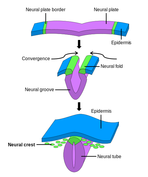
Neural Crest via Wikimedia Commons

Brain Vesicles Credit: OpenStax [CC BY 4.0] via Wikimedia Commons
Brain Divisions
The brain is the largest part of the central nervous system (CNS) and made up of three divisions: the forebrain, midbrain and hindbrain.
Forebrain
The forebrain is the largest and most complex part of the brain. It consists of the telencephalon (also known as the cerebrum or cerebral cortex), and the diencephalon. It also includes the basal ganglia as its base, which is involved in modulating motor movement.
Cerebrum
This region is involved in higher functions such as emotion, motor control, perception, personality, speech, learning and memory, thinking. It is also involved in perception of external stimuli and controlling conscious motor movement.
Hemispheres
The cerebrum is divided into right and left hemispheres. Each hemisphere generally processes sensory and motor information from contralateral limbs. Although each hemisphere has some individual responsibilities they don’t specialize in “logical” or “creative” areas and people are not left-brain or right-brain dominant.
A band of nerve fibers called the corpus callosum, along with some smaller pathways, connect the two hemispheres and enables communication between them. A corpus callosotomy (disconnecting of the two hemisphere by cutting the corpus callosum) is sometimes performed as a last ditch treatment for patients with refractory epilepsy. After this procedure, patients are left with independant hemispheres, each with their own perceptions and concepts. A split-brain patient might put down a book before they finished reading it because the hemisphere controlling the arm was not involved in reading and was ‘bored’.
Lobes and Cortices
Four lobes are used to denote specific anatomical locations and functions of specific sensory and motor hubs within the cerebrum_._ These regions process different modalities of sensation, often relayed through the spinal cord or directly through cranial nerves. In addition, one of these regions can even initiate conscious motor movement. See the table below for a description of their location and their roles.
| **Brain Lobe** | **Location** | **Role** |
| Frontal | The large frontal lobe extends from behind the forehead back to the parietal lobe.The groove in the cortex known as the central sulcus provides the border between the frontal and parietal lobes. | It is the control center for executive functions including reasoning, decision-making, expressive language, higher level cognitive processes, orientation (person, place, time, and situation integration of sensory information), and the planning and execution of movement, or motor behavior. The Frontal Lobe can be referred to as the Motor Cortex. |
| Parietal | Above the temporal lobe and adjacent to the occipital lobe, the parietal lobe houses the primary and secondary somatosensory cortex. | It plays an important role in spatial navigation and the processing of touch, pressure, temperature, and pain. |
| Occipital | The occipital lobe, located at the back of the brain. | It is the control center for the primary visual cortex, the brain region responsible for processing and interpreting visual information. |
| Temporal | The temporal lobe reaches from the temple back towards the occipital lobe. | It includes the auditory cortex and is a major processing center for receptive language. It also contains the hippocampus, a structure involved in memory formation and emotion. |
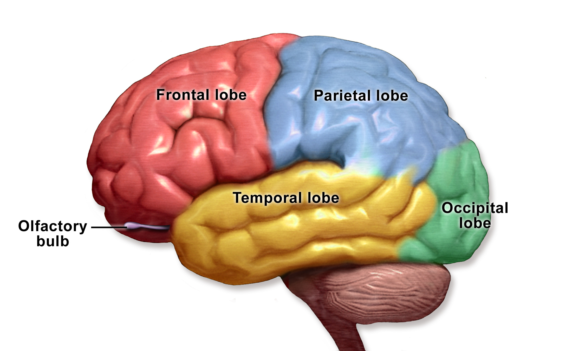
Brain Lobes Credit: Blausen.com staff (2014). “Medical gallery of Blausen Medical 2014”. WikiJournal of Medicine 1 (2). DOI:10.15347/wjm/2014.010. ISSN 2002-4436. [CC BY 3.0] via Wikimedia Commons
Detailed Topographic Mapping
Korbinian Brodmann, a German neuroscientist, first mapped the cerebrum based on its cytoarchitecture (or microstructure). He was able to subdivide the brain into 52 different Brodmann areas which, taking into account over 100 years of refining, are still used today1.

Brodmann Areas via Wikimedia Commons
Some of the areas covered by this mapping also contain their own specific regions. Both the sensory and motor cortices of the brain each have a topographic mapping to different regions of the body. This means that stimulation of a particular region of the motor cortex would produce a muscular contraction in the corresponding region. It also means that stimulation of a particular body part would activate the corresponding region in the sensory cortex (see diagram below). Similarly, the different areas of the visual cortex (Areas 17 & 18) are arranged in what is known as a retinotopic fashion. This means that different parts of this brain region relate to processing sensory information from different areas of the retina.
Topographic mapping of the sensory cortex Credit: Openstax [CC BY 4.0 US] via Openstax
Grooves
The cortex develops bumps (gyri) and grooves (sulci), enabling the brain to have more surface area and more neurons stored within the skull. No two brain cortexes are folded in the same exact way but several of these folds are large and pronounced enough to merit specific names. They are used to specify location—but also may be referred to in discussions of function.
For example, the lateral sulcus is the inner fold that separates the temporal lobe from the frontal lobe. Adjacent to the lateral sulcus is the temporal gyrus. Both this groove and fold house the primary auditory cortex, where the brain processes sound information. Wernicke’s Area, the brain region critical to processing language, also resides on the temporal gyrus.
Diencephalon
In addition to the third ventricle of the brain, the diencephalon contains a number of structures:
Hypothalamus
The hypothalamus controls activities of the autonomic nervous system, regulating neurohormones and influencing pituitary hormones, and controlling body temperature, tiredness, sleep, circadian rhythms, hunger, thirst, and behaviors related to parenting.
Thalamus
The thalamus carries sensory messages from the eyes, ears and touch receptors from all over the body. Notably, olfaction is the only sense that directly bypasses the thalamus on the way to the cortex.
Pituitary gland
The pituitary gland secretes hormones that control thyroid glands and metabolism, blood pressure, some functions of sex organs, as well as some aspects of pregnancy and growth, childbirth, nursing, water/salt concentration at the kidneys, temperature regulation, and pain relief.
Midbrain
The midbrain or mesencephalon, considered part of the brainstem, is located caudal to the forebrain, and has many different functions. Notably, neurons within the forebrain can produce the neurotransmitter dopamine. It is found in the basal ganglia, a region involved with voluntary motor action and reward based learning. Primarily, the midbrain is in charge of visual and auditory reflexes, modulating motor control, as well as sleep/wake cycles, alertness, arousal, excitation, motivation, habituation and regulation of body temperature. The human midbrain shares general architecture with the most ancient of vertebrates.
Hindbrain
The hindbrain is the most caudal part of the brain and it includes the cerebellum, pons, and medulla oblongata. Cerebellum (Latin for “little brain”) gets this name because it looks like a small version of the cerebrum. It is responsible for balance, movement, coordination and has recently been shown to be involved in some cognitive functions2. The pons has many different regions that are specialized for various functions, including: sleep, respiration, swallowing, bladder control, hearing, equilibrium, taste, eye movement, facial expressions, facial sensation, and posture. Finally, the medulla, which is also part of the brainstem, is the most caudal region of the hindbrain. It is also involved in the control of many autonomic functions, including: breathing, blood pressure, and coughing/sneezing reflexes. The cardiovascular centre, which regulates heart beats during exercise or trauma, is also located here.
Mapping the Brain in 3D
Several atlases have been developed for human brains, mouse brains, and other animal brains. Coordinate systems are used to map the size and location of each brain structure in 3D. Some systems like Talaraich coordinates use measurements relative to other prominent brain or skull features and can identify locations independent from the specific size and shape of a particular brain. These coordinate systems can be used to increase precision and replicate the effects of an experiment or surgery.
Connections and Networks
Mapping locations in the brain can help but our thoughts are more than select groups of neurons firing in a isolated regions. Modern neuroimaging research is no longer focused on the localization of function to a single area of the brain. Today, researchers are using new techniques to follow tracts of neurons that connect networks of brain areas to better understand how they work together. One technique called diffusion tensor imaging (DTI) works in combination with MRI sequences and data processing algorithms to map the diffusion of water in terms of quantity and direction of flow. It produces unparalleled images of the brain’s connected networks.
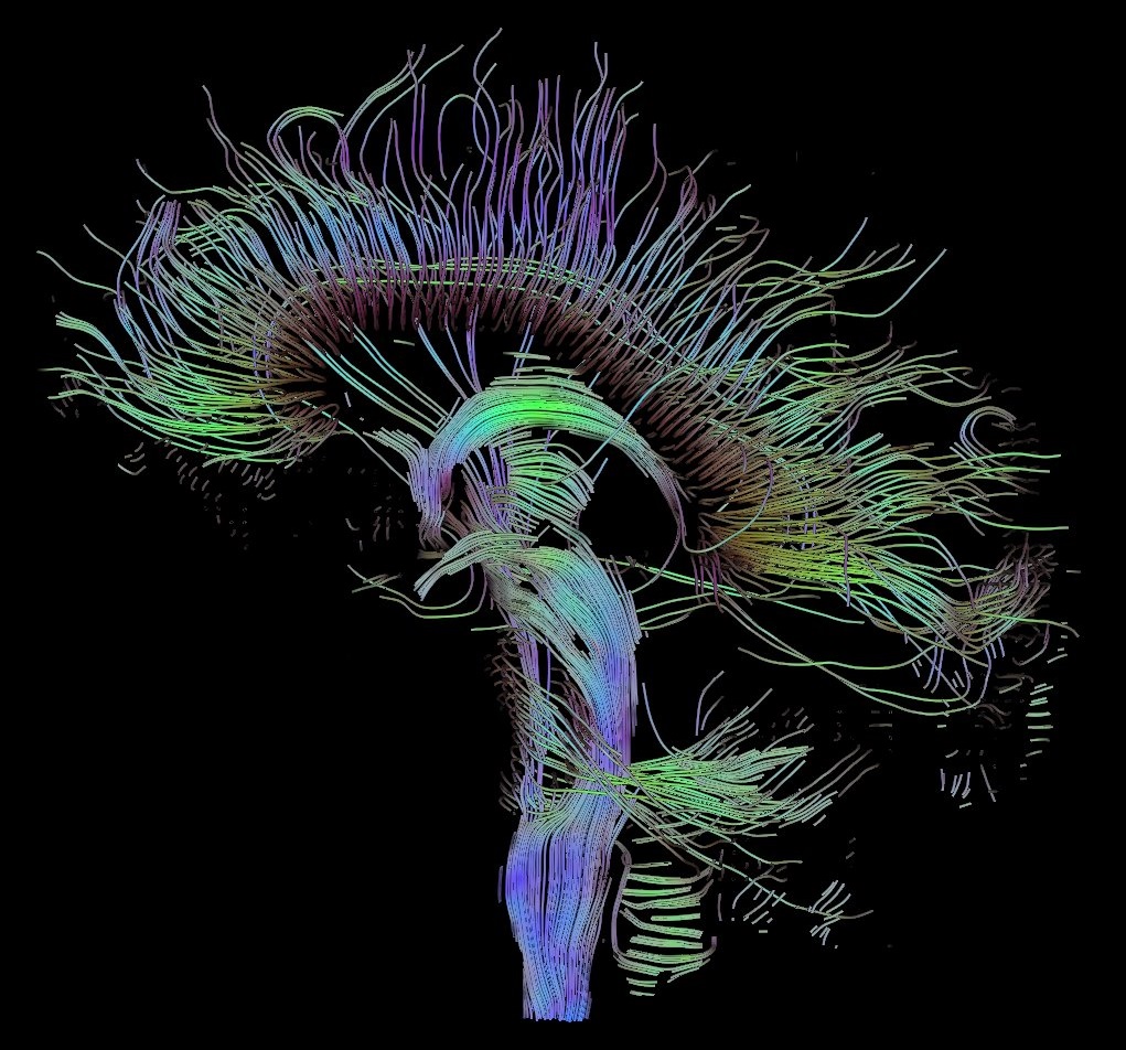
Visualization of a DTI measurement of a human brain Credit: Thomas Schultz [CC BY-SA 3.0] via Wikimedia Commons
Notes
Marios. Loukas, Christopher. Pennell, Christopher. Groat, R Shane. Tubbs, Aaron A. Cohen-Gadol; Korbinian Brodmann (1868–1918) and His Contributions to Mapping the Cerebral Cortex, Neurosurgery, Volume 68, Issue 1, 1 January 2011, Pages 6–11, ↩
Buckner, R.L., 2013. The cerebellum and cognitive function: 25 years of insight from anatomy and neuroimaging. Neuron, 80(3), pp.807-815. ↩